Rare Medical Case: Misplaced Tooth Emerges Through Teen's Eyelid
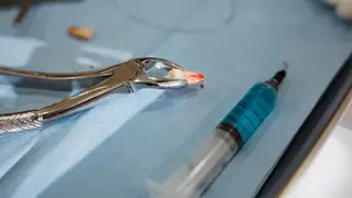
A 16-year-old boy in India presented with painless swelling on his left upper eyelid, which later developed into two hard, white projections. CT imaging revealed the projections were part of an ectopic canine tooth that had grown into the upper eye socket. Surgeons removed the tooth, and the case was reported in a medical journal.
The case involved a 16-year-old boy whose ectopic canine tooth emerged through his upper eyelid, a location never before documented in medical literature. The tooth had developed in the superior orbit, the space between eyelid and eyebrow, and was identified through CT imaging after appearing as two hard, white projections. Surgical removal confirmed it was a mature canine tooth with no signs of teratoma.
Ectopic teeth are uncommon, with roughly 2-6% of people experiencing misaligned first permanent molars within the mouth. However, teeth appearing entirely outside the dental arch are far rarer. Previously documented locations include the nasal cavity, chin, and maxillary sinus, but never the superior orbit, making this a first for medical reporting.
This case may expand clinical awareness of ectopic tooth development, potentially helping ophthalmologists and radiologists recognize unusual orbital masses during diagnosis. For patients, it underscores that rare anatomical anomalies can present with minimal symptoms, highlighting the value of imaging in puzzling presentations. The report could inform future surgical planning and differential diagnosis, though such cases remain extremely rare and unlikely to affect most individuals.