Fossilized blood vessels found inside healing T. rex rib
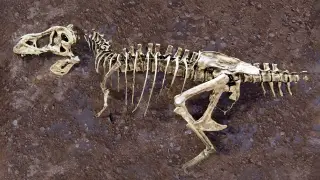
Using neutron imaging at Oak Ridge National Laboratory, researchers examined a fractured rib from Scotty, the largest known T. rex skeleton, and discovered a network of mineralized blood vessels preserved from the healing process. The dinosaur died before the fracture fully healed, and a salty marsh environment slowed decomposition, allowing the soft tissue to fossilize. This is a rare find that offers insight into dinosaur biology.
The fracture in Scotty's rib triggered a biological response that created new blood vessels, which then became mineralized over millions of years. Neutron imaging proved essential because it detects light elements like hydrogen, complementing X-ray techniques that reveal heavier elements—similar to how an MRI shows soft tissue while an X-ray shows bone density. The research began in 2020 when Mitchell, then an undergraduate, used micro-CT scanning at the Canadian Light Source to confirm fossilized soft tissue in rib sections. The specimen was discovered in Saskatchewan's Frenchman River Valley, a region rich in Late Cretaceous fossils that documents dinosaur life just before the mass extinction event.
This discovery may reshape how paleontologists approach fossil analysis, demonstrating that non-destructive imaging can reveal soft tissue structures previously thought lost to time. Researchers studying dinosaur physiology and injury recovery could gain new reference points, while the techniques showcased here may encourage broader adoption of neutron imaging across natural history collections. Public interest in T. rex discoveries could also strengthen, potentially supporting museum engagement and science education efforts around prehistoric life.