New lipid atlas identifies 539 chemical zones in the mouse brain
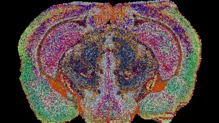
Using millions of measurements from mouse brains, researchers built the first detailed three-dimensional map of brain lipids and identified 539 distinct chemical regions. The atlas showed organized lipid patterns, including unexpected chemical variation within white matter, and may help studies of conditions such as depression and Alzheimer's disease.
Researchers at EPFL built the atlas using mass spectrometry imaging, scanning thin brain sections with a laser to detect lipids location by location. They analyzed 172 lipids across 109 slices from 11 mice, yielding about 7 million measurements, then used custom machine-learning tools to assemble a 3D reconstruction. The resulting 539 "lipizones" each carry a distinctive lipid signature.
Some chemical territories align with known anatomy or cell types, while others link distant neuron-body groups with regions connected by their fibers. Lipids may serve as shared chemical labels, giving separated but related areas a common signature. This could reveal relationships that gene-activity maps often miss.
This atlas could give neuroscientists and clinicians a new reference for studying brain chemistry, potentially aiding research into depression and Alzheimer's disease. Patients and families affected by such conditions may benefit indirectly if lipid maps help reveal disease-related changes or guide future diagnostics and treatments. However, because the work is in mice, human applications remain uncertain and likely years away.