Brain activity recordings used to reconstruct videos seen by mice
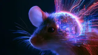
Researchers at UCL decoded signals from individual neurons in the mouse visual cortex to generate 10-second video reconstructions of what the animals viewed. This single-cell approach offers a detailed look at how visual information is represented in the brain. The method could help compare perception across species.
The reconstruction method hinged on a comparison between predicted and actual neural responses. Researchers first modeled how neurons would respond to a blank screen, then compared that baseline against real activity recorded while mice watched movies. An algorithm iteratively adjusted pixels in a blank video until the reconstruction aligned with the measured neural signals.
The team used calcium imaging to identify active individual neurons, and the encoding model also factored in mouse movements and pupil diameter changes. This single-cell approach differs from human fMRI studies, which capture broader brain signals. The researchers aim to reveal which visual features the brain emphasizes, filters, or alters before perception occurs.
This technique could eventually allow researchers to compare how different species perceive identical environments, potentially reshaping our understanding of sensory processing. If refined for human applications, it may offer new avenues for studying visual disorders or developing brain-computer interfaces. However, significant technical hurdles remain, and the method's translation from animal models to human clinical use is uncertain. The work primarily advances fundamental neuroscience rather than immediate practical applications.