Human brain organoids successfully grafted into mice after targeted tissue reduction
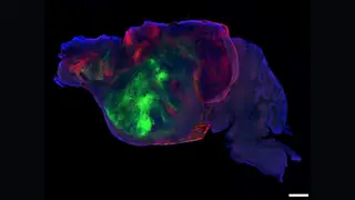
Researchers have developed a technique to shrink portions of a mouse's brain and then implant human brain organoids into the vacated space. The approach allows human neural tissue to integrate into a living rodent brain, which could enable new studies of brain development and disease. The method was described as a potential gateway for future research using human-derived tissue in animal models.
The technique involves selectively reducing portions of a mouse brain to create physical space, then implanting lab-grown human brain organoids into the vacated region. This represents a notable step in chimeric brain research, where human-derived tissue is studied within a living animal model. Previously, organoid work was largely confined to laboratory dishes, which limited insights into how neural tissue develops within a functioning, vascularized environment. By achieving integration into a living rodent brain, researchers gain a platform to observe human neural development, test disease mechanisms, and potentially screen therapeutic interventions in a more physiologically realistic context than static cultures allow.
This approach could reshape how neuroscientists study human brain disorders, offering a living model for conditions that are difficult to replicate in cell cultures. It may accelerate drug testing and deepen understanding of early neural development. However, it raises ethical questions about consciousness and the moral status of animals containing human neural tissue. Researchers, ethicists, and regulators may need to establish guidelines for how much human tissue is appropriate in animal models, balancing scientific benefit with animal welfare concerns.